Disorders of Achiles Tendon
There are many pathologic changes that may occur in the Achilles’ tendon. Tendonitis, which may be insertional (where the tendon attaches to the heel bone) or non-insertional, and acute rupture of the tendon, are 2 of the more common conditions that may require operative intervention.
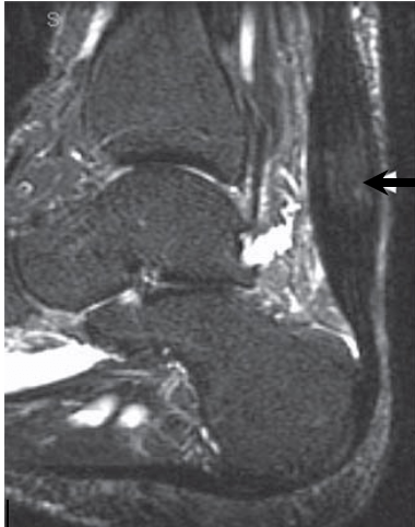
Insertional Tendonitis
Inflammation of the Achilles’ tendon where it inserts into the calcaneus (heel bone) is known as insertional tendonitis. It is often associated with an abnormal bony prominence just deep to the tendon known as Haglund’s deformity. This may play a role in rubbing on the deep aspect of the tendon to cause pain and inflammation in the tendon and surrounding soft tissues. If the associated bursitis becomes swollen enough, it can also rub on footwear or even prevent the wearing of certain shoes.
Haglund’s deformity
Non-Operative Management:
Initial management of this condition includes activity modification/rest, ice, stretching exercises, anti-inflammatories, and heel lift orthoses. A medial arch support may be of assistance by decreasing over-pronation that may exacerbate symptoms. A corticosteroid injection may help if the main problem is bursitis, but should not be given into the tendon due to increased risk of tendon rupture.
Usually the problem is manageable without surgery, but once there is a separated ossicle at the tendon insertion, surgery is usually required.
Operative Management:
The form of operative management is dependent on the degree of involvement of the Achilles’ tendon. When the tendon is not significantly involved, this condition can be managed arthroscopically. This involves 2 keyhole incisions next to the tendon through which a small camera and instrumentation can be inserted. The bursa (fluid containing sack deep to the tendon that will also be inflamed) is firstly excised, followed by the Haglund’s deformity, and finally the deep surface of the tendon is inspected. The advantage of this technique over an open (large incision) procedure is that the rehabilitation period can be accelerated.
If the Achilles’ tendon demonstrates extensive involvement on pre-operative scans or unexpectedly on arthroscopy, or if the Haglund’s deformity is too large to be safely removed by arthroscopic means, an open procedure is required. This allows removal of diseased tissue from within the Achilles’ tendon, including any ossicles and bone spurs, safe excision of a large deformity, and repair of the tendon to the calcaneal bone.
The incision for open surgery is curved and in a poor healing area. It is critical to control swelling in the early healing phase, and even then, some delayed wound healing is not uncommon.
Non-Insertional Tendonitis
Non-insertional tendonitis is inflammation due to micro tearing of some of the fibres in the Achilles’ tendon higher up the leg from the heel, typically in the middle third of the tendon. It most commonly occurs in association with running and jumping sports where forces in the Achilles’ tendon can increase to 10 times body weight. It can also occur in association with overuse syndromes, postural problems, poor footwear, or an underlying inflammatory condition that may affect multiple joints in the body.
Patients usually experience pain approximately 2-6 cm further up the leg from the heel. This may even occur at rest and/or at night. Over time, the tendon becomes thickened and an abnormal tender lump may be felt in the same area.
Non-Operative Management:
The key to successful treatment is a specific program of eccentric stretching and strengthening exercises. You will be given instruction, or be referred to a physiotherapist for this program, which is said to be successful (?75-90%?) of the time. Other management is similar to that of insertional tendonitis. Shock wave therapy may also be considered. If symptoms have gone on longer than 3 months, despite appropriate rehab, this condition becomes very difficult to manage non-operatively. When non-operative measures have failed operative management is appropriate.
Operative Management:
The idea of surgical Tenoplasty is to give the tendon a sufficient injury that the micro-tearing of the fibres can go on to heal. This is done by a small incision through which the fibres are simply split longitudinally. Initial recovery is quick, but it takes 3 months for the tendon to heal and regain strength. The success rate is very high at about 90%.
Acute Achilles’ Tendon Rupture
Rupture of the Achilles’ tendon is typically a middle-aged athletes injury, as the tendon has been shown microscopically to have suffered some very early tendonosis despite the lack of any prior discomfort. The injury is typically a rapid over-loading of an already-tensed tendon, e.g. lunging forward from a standing start, unexpected stepping in a hole, or jumping from a height. Whatever the cause, people often describe a sudden pain likened to being kicked in the back of the ankle/lower leg, which may be accompanied by an audible pop.
Non-Operative Management:
Non-operative management involves placing the leg in a below knee cast with the toes pointed. This cast is changed every 2 weeks until the ankle is finally in neutral. This process takes approximately 8 weeks.
Due to the morbidity of time on crutches and the associated high re-rupture rate (~18%), this technique is usually reserved for patients who are at high risk of complications from surgery from either an anaesthetic or surgical point of view. The latter includes people who smoke or have diabetes or vascular disease, due to their increased risk of wound breakdown and infection.
Operative Management:
An operation involves an incision to the inner side of the tendon, and the 2 tendon ends are sewn strongly back together. This makes it possible to fit a CAM (controlled ankle motion) walking boot, with the foot at the neutral position, allowing full weight bearing as soon as pain allows. The boot can be removed after the first 2 weeks for washing and active movement exercises.
Very occasionally, and especially if the repair has been particularly delayed beyond 2 weeks after injury, a strong direct repair is not possible due to significant damage in the tendon upon rupture. In this situation, a spare tendon in the back of the leg or hamstring tendon may be required to augment the repair.
Complications
A good result can be expected in 90 to 95% of patients, however no surgery is risk free. The risks and complications will be assessed and discussed with you. There is always a small risk of infection, blood clots and anaesthetic problems with lower limb surgery and measures are taken to reduce these.
Specific to the procedures described in this brochure, the area around the Achilles tendon is not a good healing area at the best of times and delayed wound healing does occur on occasion, but only rarely requires further surgery. The re-rupture rate is less than 2% and nerve damage is very uncommon (resulting in numbness of the sole or outer border of the foot.)
In any case, it is inevitable that the tendon will remain thicker than the normal tendon, and although full functional strength is to be expected, it is common that the size of the calf muscle does not come quite back to normal.
Recovery Times
Arthroscopic or Tenoplasty procedure
Hospital stay Day surgery
Rest & elevation 7 days
Crutches/frame 5-7 days
Walking boot (4-6 weeks total)
– Locked to ROM 2 weeks
– Plantarflexion 2 weeks
– Wean 2 weeks
Weight bearing
– As tolerated Day of surgery
– Full 7+days post-op
Rehabilitation (commence at…)
– ROM exercises 2 weeks
– Strength exercises 4 weeks
– Single leg calf raise 8 weeks
– Training 2-3 months
– Sport 3-4 months
Foot swelling 6-8 weeks
Time off work
– Seated 7 days
– Standing 3-4 weeks
Other Open procedures
Hospital stay 1 night
Rest & elevation 2 weeks
Crutches/frame 1-2 weeks
Walking boot (6-8 weeks total)
– Locked to ROM 2 weeks
– Plantarflexion 4 weeks
– Wean 2 weeks
Weight bearing
– As tolerated Day of surgery
– Full 2+weeks post-op
Rehabilitation (commence at…)
– ROM exercises 2 weeks
– Strength exercises 6-8 weeks
– Single leg calf raise 10-12 weeks
